OCT Technology

Principle of OCT Technology

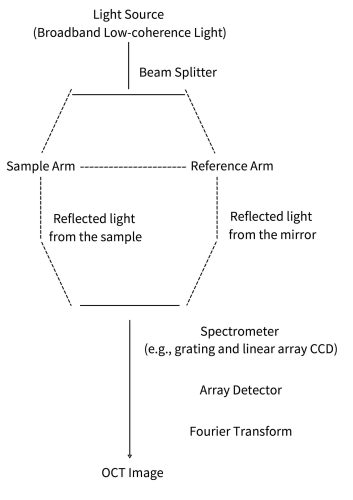
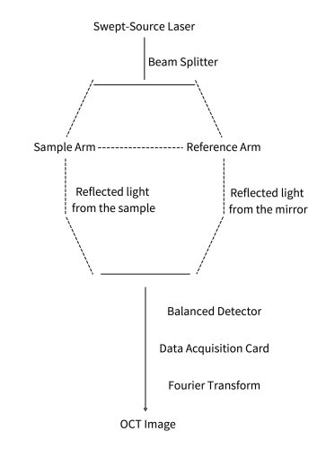
Optical Coherence Tomography (OCT) is a non-invasive, high-resolution imaging technique widely used in medical diagnosis, industrial inspection and other fields.
Its core principle is based on low-coherence interferometry, which combines the interference characteristics of light waves to reconstruct 2D or 3D images of the internal structure of samples.

Basic Principles

Optical Coherence Tomography (OCT) is an advanced imaging technology based on low-coherence optical interferometry. Light from a broadband source is divided into two paths: a sample beam, which illuminates the target and undergoes backscattering within the tissue or material, and a reference beam, which reflectsfrom a fixed reference mirror. The two beams are then recombined to produce aninterference signal.
Interference occurs only when the optical path difference between the sample and reference arms lies within the coherence length of the light source. By analyzing changes in this interference signal as the optical path length varies, OCT extracts depth-resolved structural information. This enables high-resolution cross-sec-tional (2D) and volumetric (3D) imaging of internal microstructures - non-invasively and in real time.

OCT原理英文

Specific Implementation Method

Light Source

Broadband light sources such as superluminescent diodes (SLEDs) or broadband lasers are typically used. The emitted light has a wide spectral range to achieve high-resolution depth measurement.

Detector

High-speed photodetectors are used to detect interference signals. The detector converts optical signals into electrical signals and records information such as the intensity and phase of the interference signals.

Optical Interferometer

A Michelson interferometer structure is generally adopted, which splits the incident light into a sample beam and a reference beam, and causes the two beams to interfere when returning. By precisely controlling the length of the reference arm, the optical path difference can be adjusted to scan sample information at different depths.

Signal Processing and Image Reconstruction

A computer is used to process and analyze the massive interference signals collected by the detector. Through algorithms such as Fourier transform, the interference signals are converted from the time domain to the frequency domain, thereby obtaining the reflectivity information of the sample at different depths. Based on this information, appropriate image reconstruction algorithms, such as those based on raster scanning or line scanning, are employed to construct two-dimensional or three-dimensional images of the sample.

SD-OCT and SS-OCT

SD-OCT

Spectral-Domain OCT (SD-OCT) implements frequency-domain analysis through a spectrometer and array detector, and is suitable for static or medium-speed high-resolution imaging, such as ophthalmic retinal imaging.

SS-OCT

Swept-Source OCT (SS-OCT) utilizes a swept-source laser and a single-point detector to capture deep tissue information at ultra-high speed. It is suitable for dynamic scenarios (such as cardiovascular blood flow) and industrial online inspection.

Key Parameters of OCT

Axial Resolution

Definition: The resolving capability along the direction of light propagation, typically 1–15 micrometers.

Determining factor: Bandwidth of the light source (the wider the bandwidth, the higher the resolution).

  • Δz: Axial resolution (Unit: m, usually expressed in μm)
  • λ0​: Central wavelength of the light source (Unit: m)
  • Δλ: Spectral bandwidth of the light source (Unit: m)

Lateral Resolution

Definition: The resolving capability perpendicular to the direction of light propagation, typically 5–20 micrometers.

Determining factors: Numerical aperture (NA) of the objective lens and the focused spot size, similar to conventional microscopy.

  • Δx: Lateral resolution (Unit: m, usually expressed in μm)
  • λ0​: Central wavelength of the light source (Unit: m)
  • NA: Numerical Aperture of the objective lens, defined as NA=nsinθ
  • n: Refractive index of the medium between the objective lens and the sample (n=1 in air, n≈1.33−1.4 in tissue or water)
  • θ: Half-angle of the objective lens aperture (half-angle of the focused light cone)

Light Source Parameters

Center Wavelength:

  • 800~900 nm: Suitable for shallow-layer high-resolution imaging (e.g., retina, high-resolution surface inspection, transparent/translucent materials, etc.).
  • 1300~1550 nm: Suitable for deep tissue imaging (e.g., skin, blood vessels, composite materials, silicon-based materials, thick materials, highly scattering materials, etc.).

Bandwidth: Determines the axial resolution. Superluminescent diodes (SLD) or supercontinuum light sources can achieve broadband spectra.

Imaging Depth

Definition: The maximum depth that light can penetrate into tissue, typically 1–3 mm (depending on tissue type).
Influencing factors: Source wavelength (e.g., 1300 nm penetrates deeper than 800 nm but has higher scattering), and tissue scattering properties.

Scanning Speed

Definition: The number of A-scans (axial scans) acquired per second.
Frequency-domain OCT can achieve tens of thousands to hundreds of thousands of A-scans per second, making it suitable for imaging dynamic tissues such as the heart and blood flow.