News Information
Clairvo Technology has been honored with the title of "High-tech Enterprise", and its technological innovation capabilities have once again been recognized by the national authorities
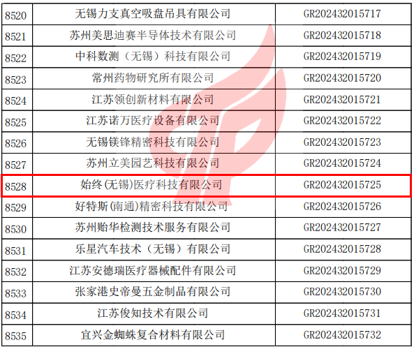
On January 7, 2025, the National Leading Group Office for the Administration of High-tech Enterprise Certification issued the “Announcement on the Filing of the Third Batch of High-tech Enterprises Recognized and Filed by the Certification Body of Jiangsu Province in 2024”. Clairvo Technology was on the list and officially approved as a national high-tech enterprise. This marks that Clairvo Technology has entered the ranks of national high-tech enterprises in terms of comprehensive indicators such as core independent intellectual property rights, the ability to transform scientific and technological achievements, the level of R&D organization and management, and the growth potential of the enterprise.
The recognition of National High-tech Enterprises is jointly implemented by the Ministry of Science and Technology, the Ministry of Finance and the State Taxation Administration. It is one of the most valuable honors for technological innovation enterprises in China. Since its establishment, Clairvo Technology has been dedicated to the independent research and development and industrial application of optical coherence tomography (OCT) technology. It has now formed a multi-dimensional product matrix covering fields such as medical precision diagnosis and treatment and industrial non-destructive testing. It has cumulatively applied for and obtained more than 20 patents and software Copyrights. The performance indicators of its core products have reached the international advanced level, successfully achieving import substitution.
This recognition as a high-tech enterprise not only highly affirms the company’s technological research and development strength and innovation capabilities, but also provides strong policy support for the company in undertaking national science and technology plan projects and attracting high-end talents. In the future, Clairvo Technology will take this as a new starting point, continuously increase investment in research and development, accelerate the layout of cutting-edge technologies such as multimodal OCT and AI intelligent image analysis, further consolidate and expand its leading position in the domestic and international high-end medical and industrial inspection markets, and contribute “Always” strength to the high-quality development of China’s high-end optical imaging industry.