Fully Automatic
High Speed Swept OCT
-1-927x1200.png)
Clairvo One
Clairvo One is the first full-eye OCT ophthalmic testing device pioneered by Clairvo Medical. Adopting the most advanced swept-source OCT imaging technology combined with an innovative optical path design, the device features stronger penetration, clearer detection and high-precision imaging quality. It also integrates multiple cameras, collaborative robots and machine vision algorithms to achieve a fully automatic autofocus function. The testing system is capable of completing nearly all ophthalmic examinations including anterior and posterior segment OCT, OCTA and visual biometry. Equipped with an AI-assisted image interpretation system, it can reduce the testing time by 90%, delivering greater value to hospitals, physical examination institutions and patients alike.
Six Characteristics
Innovative Structural Design
Fully automatic robotic arm control design, adaptive tracking and shooting, one-click sample acquisition
High-Speed Scanning Rate
A scanning rate of 400,000 A-scans per second,delivering ultra-high resolution and precise tomographic information of the entire eye.
Intelligent Analysis System
AI-assisted intelligent image analysis system,enabling efficient screening of fundus lesions and empowering medical diagnosis.
Superb PenetrationPerformance
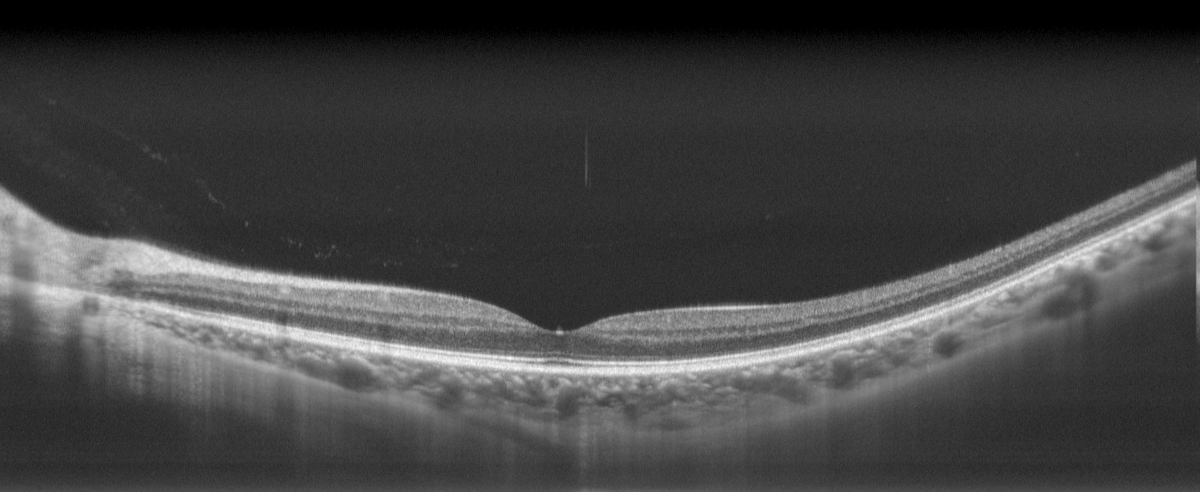
Excellent tomographic capability for easier penetration of the fundus,realizing the visualization of choroidal and scleral tissue information.
Larger Imaging Depth
Flexible adjustment of imaging depth,facilitating and enabling anterior segment imaging, large-range fundus imaging and axial length imaging.
Clear Blood Flow Imaging
Capturing high-definition OCTA blood flow imaging results with higher quality,and detecting neovascularization in the choroidal capillary layer.
Image Generation
It greatly improves the efficiency of fundus imaging data acquisition and optimizes the acquisition operation process.
Indications
Macular Diseases
OCT examination can identify the presence of neovascularization, defects or hyper-reflective areas in each tomographic layer—these abnormalities in various retinal regions are all causative factors of macular diseases.
Diabetic Retinopathy
Typical fundus changes at all stages of diabetes can be visualized on OCT, such as punctate exudates in the fundus, detachment of the neuroepithelial layer at the fovea centralis, and retinal hemorrhage.
Cataract
OCT is also a mandatory examination item for cataracts both preoperatively and postoperatively: it provides guiding references for surgical planning and enables postoperative evaluation of the patient’s fundus condition.
Corneal Lesions
It allows for non-contact assessment of corneal status, including corneal thickness, the location of corneal lesions, and corneal morphology.
Optic Nerve Diseases
OCT can reveal pathological changes that are difficult to detect in conditions such as glaucoma, including varying degrees of thinning of the peripapillary nerve fiber layer and thinning of the optic nerve fiber layer.
High Myopia
OCT enables the assessment of the posterior pole of the retina for atrophic thinning, retinal schisis, shallow retinal detachment, and vitreomacular traction, among other abnormalities.
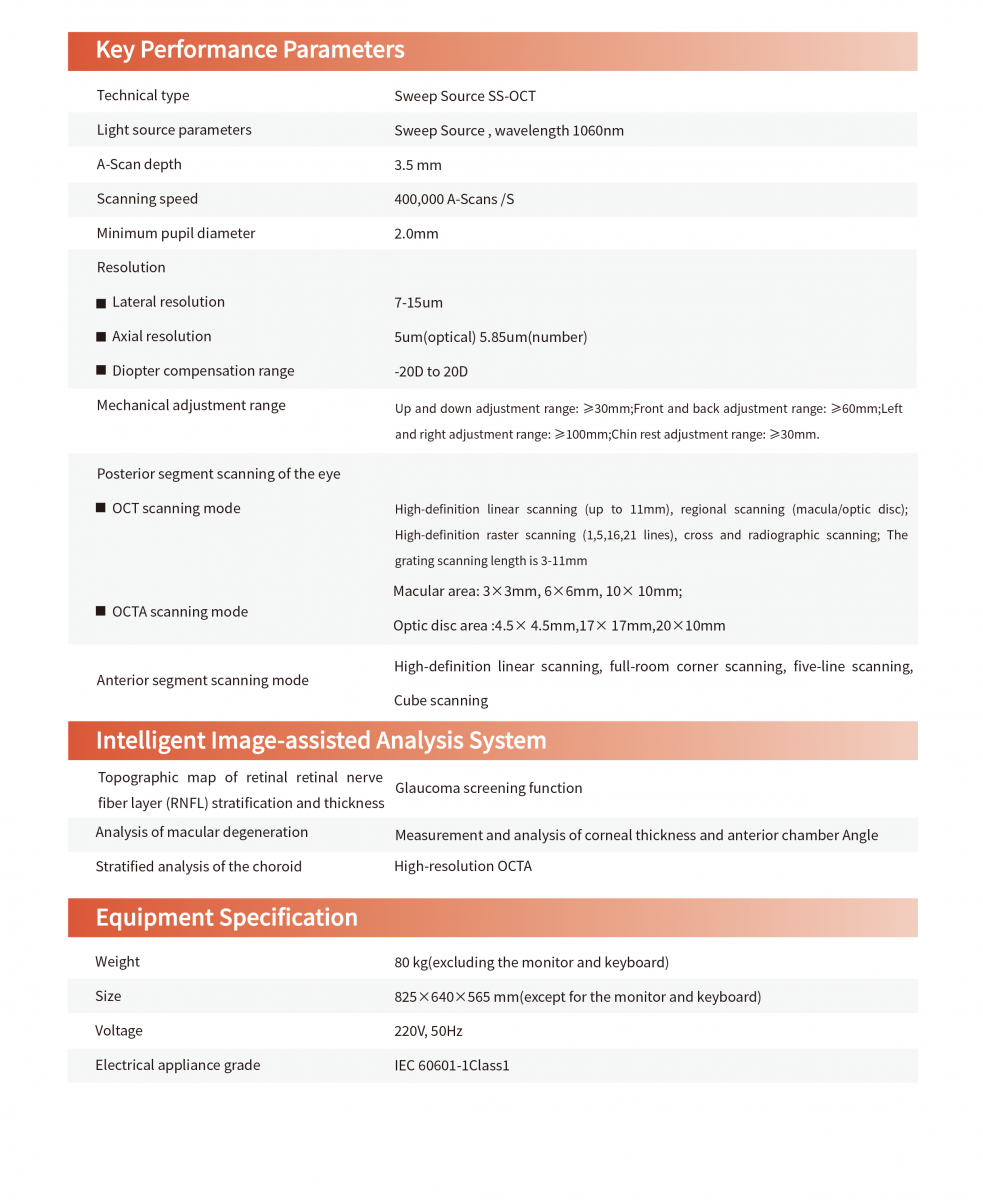
Technical Parameters Of Ophthalmic OCT