Optical Coherence Tomography (OCT) is a non-invasive, high-resolution imaging technology widely used in medicine, industrial inspection and other fields. Its core principle is based on low-coherence interferometry, which combines the interference characteristics of light waves to reconstruct 2D or 3D images of the internal structure of a sample.
Basic Principle
OCT utilizes the principle of optical low-coherence interferometry. It splits the light emitted by a broadband light source into two beams: one beam irradiates the measured object (sample light), and the light undergoes backscattering after entering the measured object. The other beam acts as reference light, which is reflected back by a fixed reference mirror. The two beams recombine at the beam splitter to generate an interference signal. An effective interference signal is only formed when the optical path difference (OPD) between the two arms is within the coherence length of the light source. By measuring the variation of the interference signal with the optical path difference, information about different depths inside the sample can be obtained, and then a 2D or 3D structural image of the sample can be reconstructed.
Specific Implementation Methods
Light Source
Broadband light sources such as Superluminescent Light-Emitting Diodes (SLED) or broadband lasers are typically adopted, which emit light with a wide spectral range to achieve high-resolution depth measurement.
Detector
A high-speed photodetector is used to detect interference signals. The detector converts optical signals into electrical signals and records information such as the intensity and phase of the interference signals.
Optical Interferometer
A Michelson interferometer structure is generally used, which splits the incident light into sample light and reference light and enables the two beams to interfere upon their return. By precisely controlling the length of the reference arm, the optical path difference (OPD) can be adjusted to scan sample information at different depths.
Signal Processing and Image Reconstruction
A computer is used to process and analyze the large number of interference signals collected by the detector. Through algorithms such as Fourier transform, the interference signals are converted from the time domain to the frequency domain, thereby obtaining the reflectivity information of the sample at different depths. Based on this information, appropriate image reconstruction algorithms (e.g., raster scanning or line scanning-based methods) are adopted to construct 2D or 3D images of the sample.
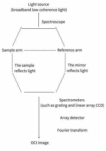
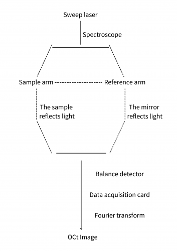
SD-OCT and SS-OCT
SD-OCT
Spectral Domain OCT (SD-OCT) achieves frequency domain analysis via a spectrometer and an array detector, making it suitable for static or medium-speed, high-resolution imaging scenarios such as ophthalmic retinal imaging.
SS-OCT
Swept-Source OCT (SS-OCT) utilizes a swept-source laser and a single-point detector to acquire deep tissue information at an ultra-high speed. It is applicable for dynamic scenarios (e.g., cardiovascular blood flow) and industrial online inspection.
Key Parameters of OCT
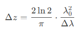
Axial Resolution
Definition: Resolution capability along the light propagation direction, typically ranging from 1 to 15 μm.Determining Factor: Spectral bandwidth of the light source (the wider the bandwidth, the higher the resolution).
Δz: Axial resolution (Unit: m, usually expressed in μm)
λ0: Central wavelength of the light source (Unit: m)
Δλ: Spectral bandwidth of the light source (Unit: m)
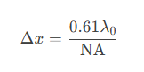
Lateral Resolution
Definition: Resolution capability perpendicular to the light propagation direction, typically ranging from 5 to 20 μm.Determining Factor: Numerical Aperture (NA) of the objective lens and the size of the focused light spot, consistent with the principle of traditional microscopes.
Δx: Lateral resolution (Unit: m, usually expressed in μm)
λ0: Central wavelength of the light source (Unit: m)
NA: Numerical Aperture of the objective lens, defined as NA=nsinθ
n: Refractive index of the medium between the objective lens and the sample (n=1 in air, n≈1.33−1.4 in tissue or water)
θ: Half-angle of the objective lens aperture (half-angle of the focused light cone)
1300~1550 nm: Suitable for deep tissue/ material imaging (e.g., skin, blood vessels, composite materials, silicon-based materials, thick materials, highly scattering materials, etc.).
Spectral Bandwidth:Determines the axial resolution; broadband spectra can be achieved by superluminescent diodes (SLD) or supercontinuum light sources.
Imaging Depth
Definition: The maximum penetration depth of light in the tissue/material, typically 1~3 mm (dependent on the tissue/material type).Influencing Factors: Central wavelength of the light source (e.g., 1300 nm light penetrates deeper than 800 nm light but with stronger scattering), and scattering characteristics of the tissue/material.
Scanning Speed
Definition: The number of A-scans (axial scans) acquired per second.Frequency domain OCT can achieve tens of thousands to hundreds of thousands of A-scans per second, making it suitable for imaging dynamic tissues (e.g., heart, blood flow).