Fully Automatic
High Speed Sweep OCT
-1-927x1200.png)
Clairvo One
Clairvo One is a whole-eye OCT ophthalmic testing device pioneered by Clairvo Medical. The equipment adopts the most advanced sweep frequency light source OCT imaging technology and innovative optical path design, which has stronger penetration, clearer detection and high-precision imaging quality. At the same time, it also uses multiple cameras, collaborative robots and machine vision algorithms to achieve fully automatic self-focusing function, and the detection system can complete almost all eye detection items such as front and back node OCT, OCTA, and visual biometrics. The AI-assisted film reading system can shorten the test time by 90%, creating higher value for hospitals, medical institutions and patients.
Six Characteristics
Innovative structure design
Automatic robotic arm control design, adaptive tracking shooting, one-click pattern book collection.
High scanning rate
The scan rate of 400,000 times per second provides ultra-high resolution accurate tomography information for the whole eye.
Intelligent analysis system
Intelligent image-assisted analysis system can realize efficient fundus lesion screening and enable medical diagnosis.
Super penetrating effect
Good chromatographic ability, easier to penetrate the fundus, to achieve the display of choroidal and scleral tissue information.
Greater imaging depth
Flexibly realize the change of imaging depth, providing convenience and feasibility for anterior segment imaging, wide-range fundus imaging, and axial image development.
Clear flow imaging
Better access to high-resolution OCTA blood flow imaging results and detection of neovascularization in the choroidal capillary layer.
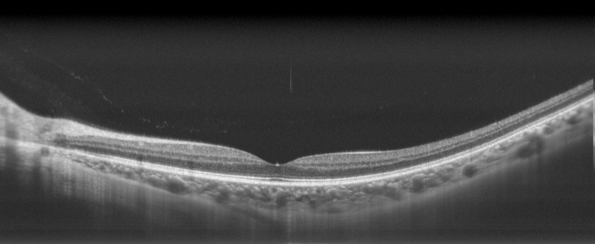
Image Generation
Greatly improve the efficiency of fundus imaging data acquisition and optimize the acquisition operation process.
Applicable Disease
Macular disease
OCT examination shows that each section has new blood vessels, defects, or hyperreflective areas, all of which can cause macular disease.
Diabetic retinopathy
The typical fundus changes at all stages of diabetes can be shown on OCT. Such as fundus spot exudation, macular fovea nerve cortex detachment, bleeding and so on.
Cataract
OCT is also a must-check item before and after cataract surgery, which has a guiding role in surgery and can evaluate the fundus condition of patients after surgery.
Corneal lesions
The corneal condition can be viewed, such as corneal thickness, corneal lesion location, corneal shape, etc., and OCT does not touch the cornea.
Optic nerve disease
OCT can show different degrees of thinning of the nerve fiber layer around the optic disc, thinning of the optic nerve fiber layer, and pathological changes such as glaucoma cup that cannot or cannot be easily detected by routine examination.
High myopia
Through OCT, we can know whether the posterior polar retina is atrophic and thin, whether there is retinal cleft and shallow retinal detachment, and whether there is vitreous macular traction.
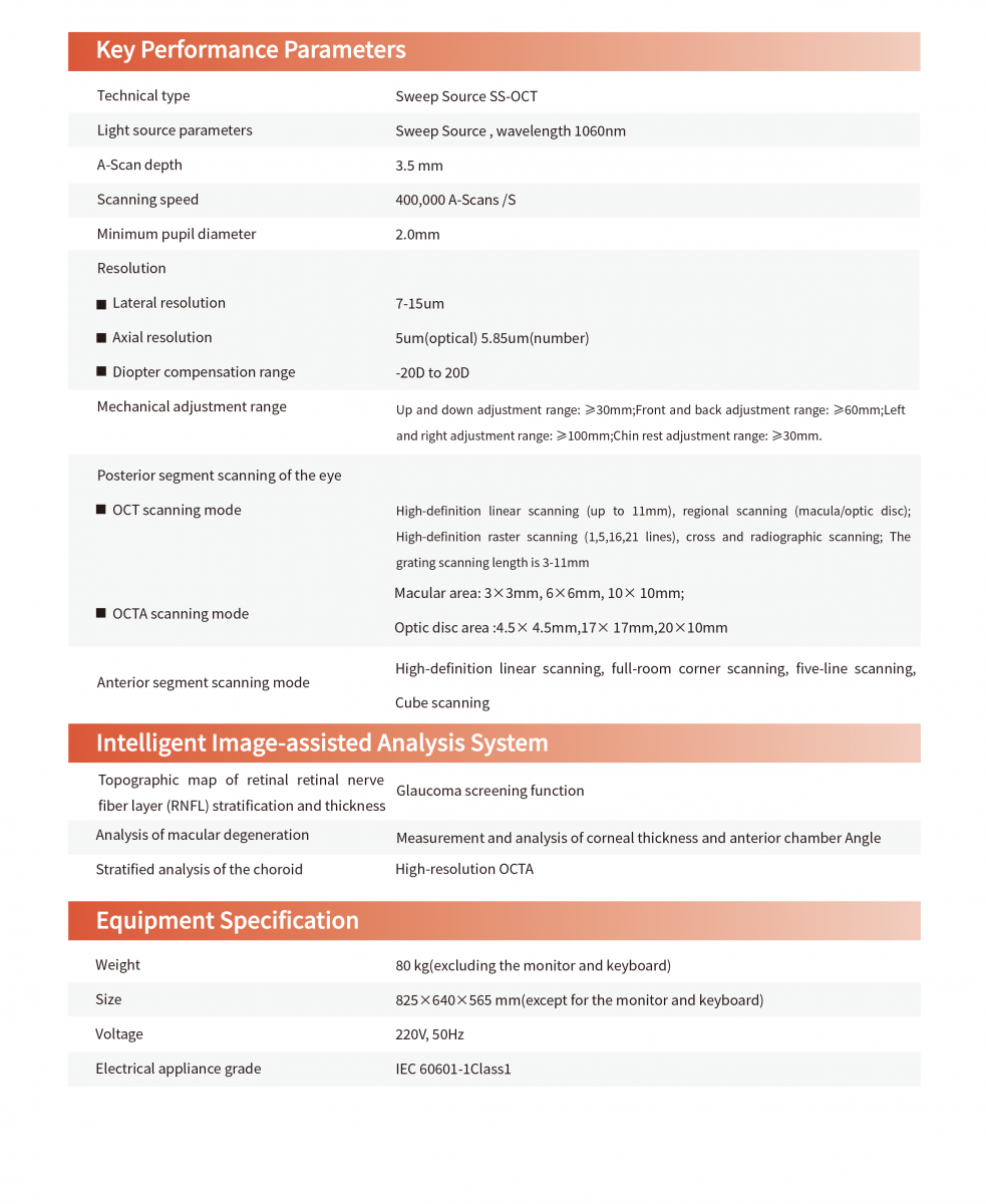
Technical Parameters Of Ophthalmic OCT