Skin cancer is the most commonly occurring cancer in humans, with an incidence that has steadily increased worldwide. The prognosis of advanced melanoma is poor, and carcinomas are associated with high morbidity.
60% biopsies result in benign diagnoses
Although dermoscopy is a useful tool for clinical examination, the sensitivity of dermoscopic monitoring is limited by melanomas that may arise in normal skin or in clinically benign nevi that were not initially photographed.
20% skin cancers missed at early stage
Dermoscopy improves the diagnostic accuracy for melanoma but only for experienced examiners. Dermoscopy by untrained or less experienced examiners is no better than clinical inspection without dermoscopy.
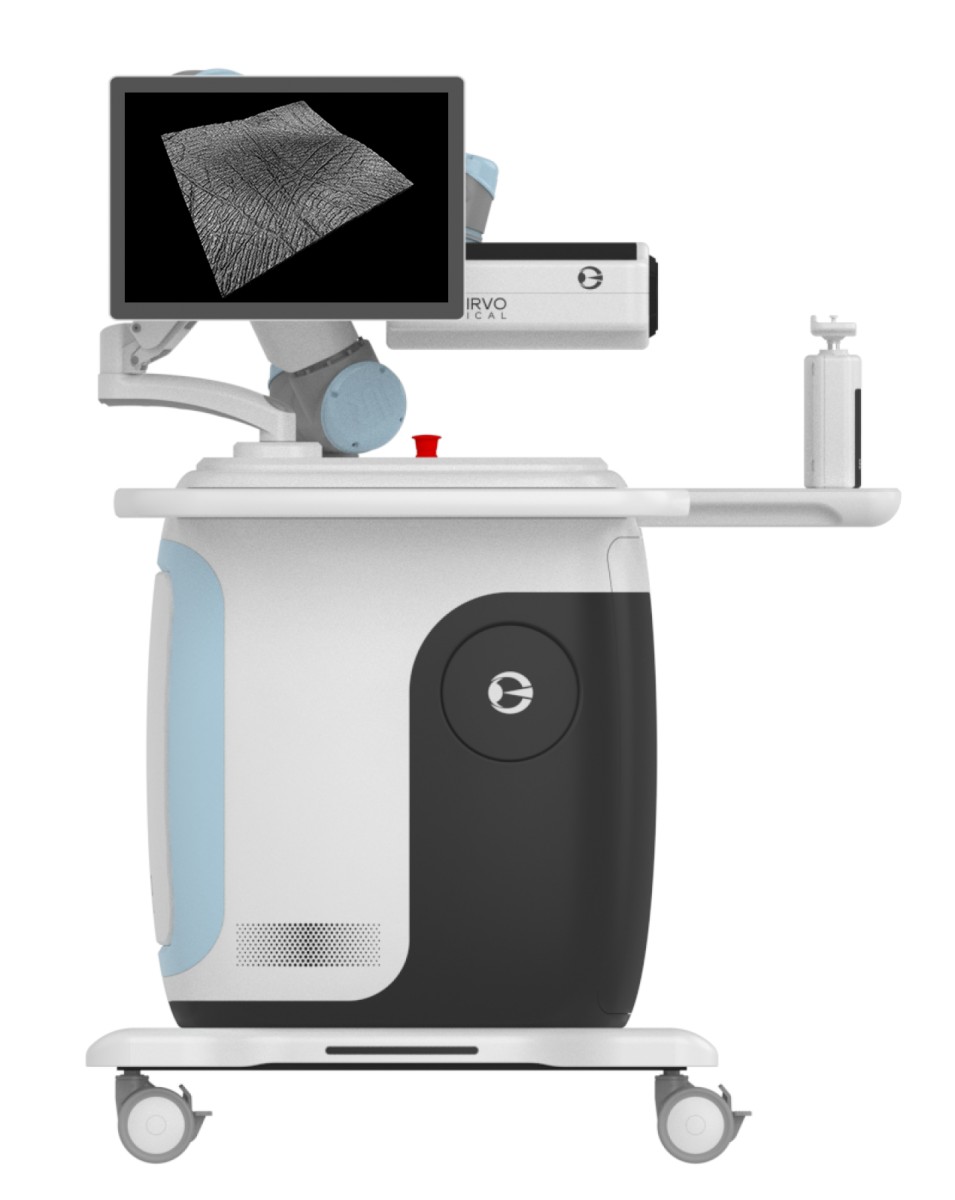
Image Generation
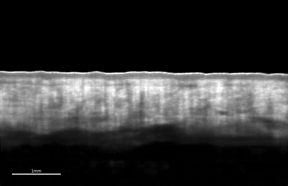
OCT vertical (top left), horizontal (bottom left) images and 3D stack (right) of healthy human skin in vivo.
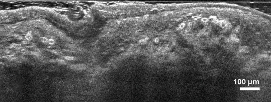
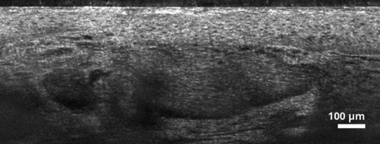
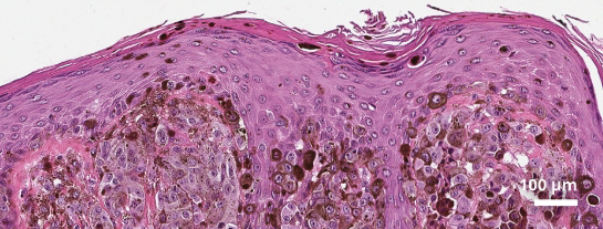
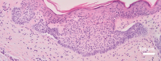
OCT vertical images of a melanoma left) and a superficial basal cell carcinoma (right) with corresponding H&E histopathology images.
For damaged skin, longitudinal cross-sectional images can be quickly collected to reveal the substructure of the dermis, from which a 3D volume or in-plane image of the entire wound can be generated.
Significant depth visualization of the structure and vascular system enables clear observation of the physiological structures of the stratum corneum, hyaline layer, epidermodermal junction and dermal tissue.
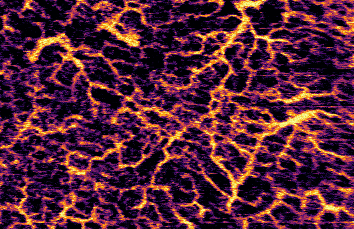
It can provide more information about the vascular network. Not only small blood vessels located in the superficial layer were observed, but also large blood vessels penetrating the deep dermis.
If you want more information,
Please click the button below "Learn more"