News Information
Clairvo Medical Technology Co., Ltd. has been successfully selected as a technology-based small and medium-sized enterprise, and its innovation-driven development has once again been recognized at the national level
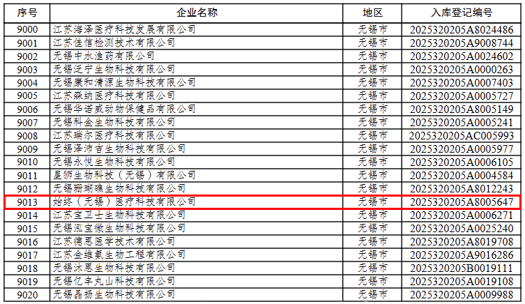
On September 5, 2025, the Department of Science and Technology of Jiangsu Province issued the “Announcement on the First Batch of Technology-based Small and Medium-sized Enterprises in Jiangsu Province in 2025”. With its outstanding performance in core technologies such as optical coherence tomography (OCT), product innovation and R&D management, Clairvo Technology was successfully included in the national technology-based small and medium-sized enterprise database.
This inclusion in the database marks that Clairvo Technology’s independent innovation capabilities and the level of scientific and technological achievement transformation in the fields of high-end medical imaging and industrial inspection have been recognized by the national authorities. Since its establishment, the company has always adhered to the R&D route of “independent control and domestic substitution”, and has applied for/been granted more than 20 invention patents, utility model patents and software Copyrights. Its core OCT products are widely used in scenarios such as precise ophthalmic diagnosis and treatment, non-destructive testing of semiconductors, and material science analysis.
In the future, Clairvo Technology will rely on the policy support for technology-based small and medium-sized enterprises, continuously increase investment in research and development, accelerate the next-generation upgrade of multimodal OCT and AI intelligent analysis platforms, further expand the dual-wheel drive market of medical and industrial sectors, build a benchmark enterprise for high-end optical imaging in China, and contribute technological strength to the “Healthy China” and “Quality Strong Country” strategies.